
The order of the pseudoscorpions (Pseudoscorpiones)
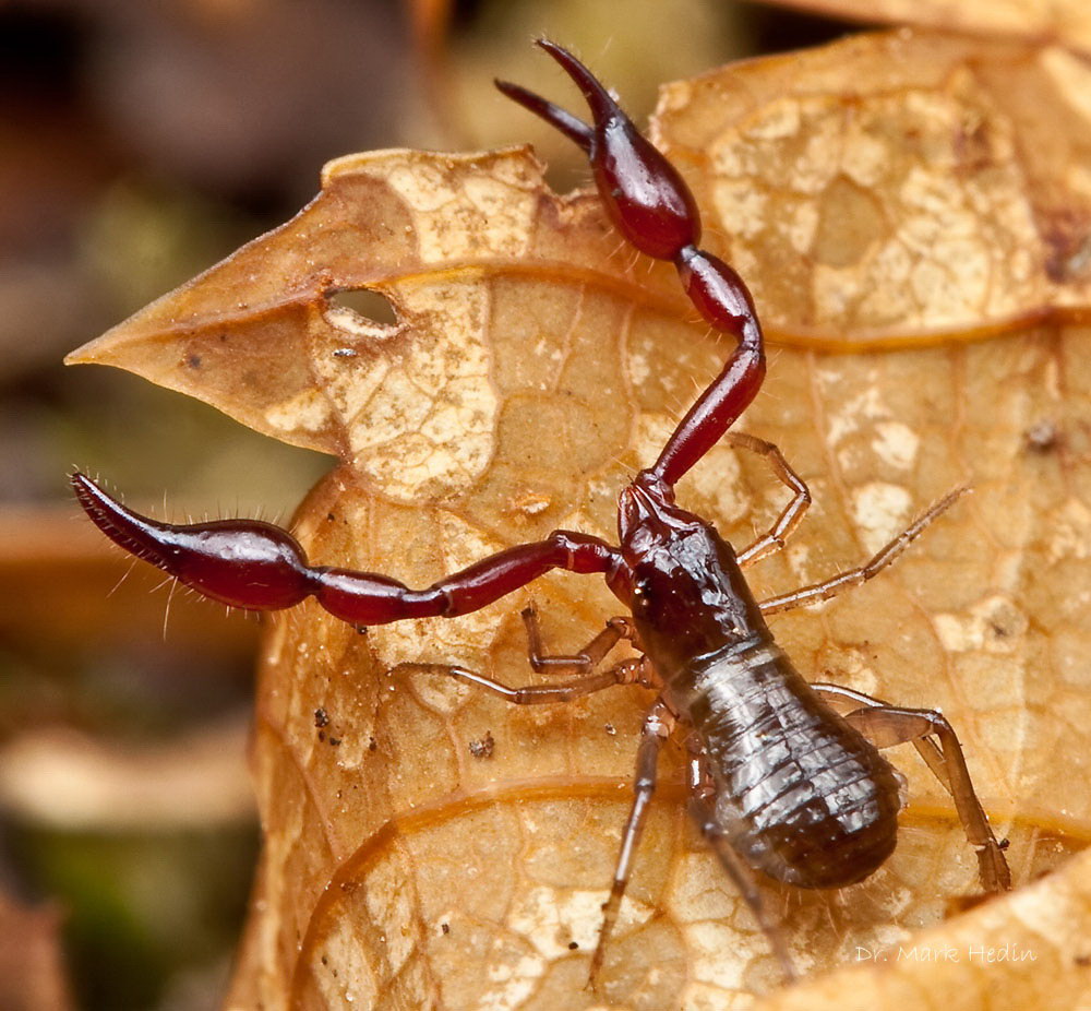
Figure 1: Pseudoscorpion of the suborder Neobisioidea. The picture was taken at a Tsuga-population at St. Joe National Forest, Idaho. Pseudoscorpions of the suborder Neobisioidea possess venom glands within their pedipalpal chelae and therefore belong to the Iocheirata. (Harvey, 1992; Murienne et al., 2008; Weygoldt, 1969, p5). This species possesses two pairs of lateral eyes. Copyright is held by Dr. Mark Hedin, website: http://marshalhedinlab.com/, photostream: http://www.flickr.com/photos/23660854@N07/.
The pseudoscorpions consisting of 3385 described species (Harvey, 2007; Harvey, 2009) represent a meso-diverse order within the arachnids (Harvey, 2002). Pseudoscorpions are terrestrial predators, that superficially resemble scorpions devoid of a tail and a stinger (Harvey, 2002). Their body sizes range between 1 mm up to more than 10 mm (Harvey, 2002; Murienne et al., 2008). The oldest pseudoscorpions are known from the Devonian (380 mio. years before present) (Schawaller et al., 1991).
Most of the pseudoscorpion species occur in the tropics and sub-tropics though they are also to find in temperate zones and their distribution expands far in high northern
and southern latitudes. The species Neobisium muscorum occurs in Scandinavia (Weygoldt, 1969, p108).
Most of the species are restricted in their distribution, however some species like Chelifer cancroides and Cheiridium museorum are cosmopolitan (Weygoldt, 1969, p108).
An essential factor for the occurrence of pseudoscorpions is the presence of small cracks and slits in the environment into which the animals can retreat (Weygoldt, 1969, p108).
Many species occur in the leaf litter of woods, where especially representatives of the families Chthoniidae, Neobisiidae and Chernetidae can be found.
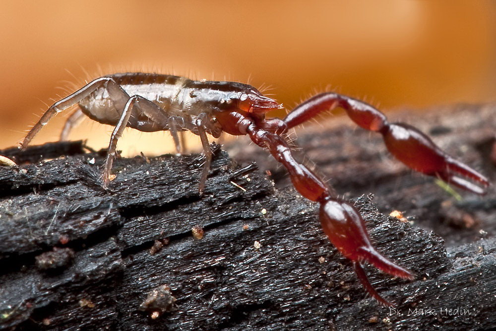
Figure 2: Pseudoscorpion of the suborder Neobisioidea. The picture was taken at a Tsuga-population at St. Joe National Forest, Idaho. Pseudoscorpions of the suborder Neobisioidea possess venom glands within their pedipalpal chelae and therefore belong to the Iocheirata. (Harvey, 1992; Murienne et al., 2008; Weygoldt, 1969, p5). This species possesses two pairs of lateral eyes. Copyright is held by Dr. Mark Hedin, website: http://marshalhedinlab.com/, photostream: http://www.flickr.com/photos/23660854@N07/.
Another important habitat of pseudoscorpions is the bark of trees under which representatives of the families Chernetidae and Cheliferidae and in North America of the families Sternophoridae, Olpidae and Atemnidae can be found.
Cracks in rock serve also as living environment and some species are pure cave dwellers (Fig. 3, 5) with specific morphologic adaptations like highly elongated
appendages and the lack of eyes (mainly representatives of the families Chthoniidae and Neobisiidae) (Weygoldt, 1969, p109-110).
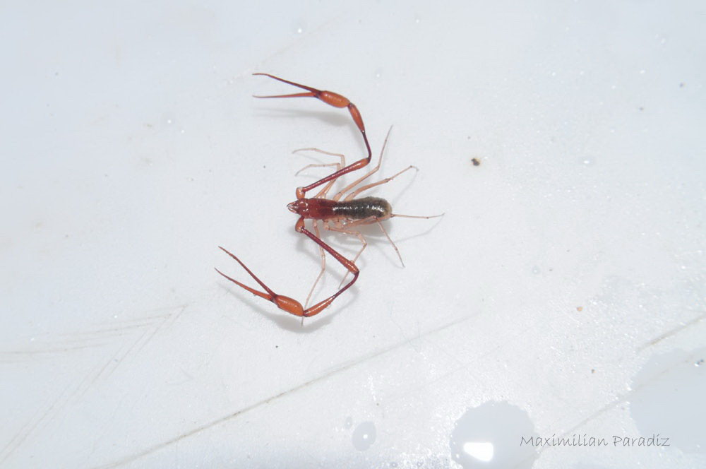
Figure 3: Vachonium cryptum of the family Bochicidae. Collected in the caves of Calcehtok, Yucatan, Mexiko. An iocheirat troglomorph pseudoscorpion whose venom glands of the fixed pedipalpal finger are highly reduced (Murienne et al., 2008). The pedipalps and walking legs of this pseudoscorpion are highly elongated which is often a result of an adaptation to a life as cave dweller (Weygoldt, 1969, p109-110). Copyright is held by Maximilian Paradiz.
Even the intertidal zones at the seashore, which are submerged twice a day, are inhabited by some species (for example Neobisium maritimum). Nests of birds and small mammals are habitats of further species (Weygoldt, 1969, p112). A certain amount of species inhabits buildings (Weygoldt, 1969, p109).
The body of pseudoscorpions is divided into the tagmata pro- and opisthosoma (Weygoldt, 1969, p3).
The ventral prosomal surface is not covered by sternal plates (in some families a rudimentary sternum can be found) but rather by the coxae or additional basal segments of the walking legs (Weygoldt, 1969, p3).
The chelicerae of the pseudoscorpions consist of two-segmented chelae that display a galea/spinneret on the tip of the movable cheliceral finger, which contains the
opening of the efferent duct of the silk glands (Weygoldt, 1969, p4). The occurrence of silk glands whose efferent ducts open in the chelicerae is a unique feature of pseudoscorpions (Murienne et al., 2008).
The pedipalps of pseudoscorpions bear large chelae that resemble those of scorpions. They constitute the most important sensoric organs of pseudoscorpions and are utilized for prey manipulation and smaller particles for nest building as well as social interactions like inner specific fights and mating (Weygoldt, 1969, p4).
The tips of the chelal fingers are bent inward and in pseudoscorpions belonging to the group of the Iocheirata (“poison-handed”) there can be found the openings of the efferent duct of the venom glands (Harvey, 1992; Murienne et al., 2008; Weygoldt, 1969, p5).
Venom glands and the openings of their efferent ducts can occur in one or both fingers of the chelae (Harvey, 1992; Murienne et al., 2008; Weygoldt, 1969, p15).
The number of segments of the walking legs is reduced to 6 or 5 instead of the original 7 segments (Weygoldt, 1969, p5). The tarsi bear arolia/pulvilli which allow the pseudoscorpions to walk on smooth surfaces (Weygoldt, 1969, p5).
When touched posteriorely pseudoscorpions immediately turn around and present their chelae to the threat. Their escape is faciliated by the ability to walk faster backwards than forward (Weygoldt, 1969, p22).
The junction between pro- and opisthosoma is broad by what there is no mobility between these tagmata (except for the Feallidae) (Weygoldt, 1969, p6).
The opisthosoma is segmented and consists of 12 segments (Weygoldt, 1969, p6).
The tergits can be uniform (Fig. 1-5) as it is the case in the Chthoniidae, Neobisiidae and others or divided (Fig. 6, 7) in two lateral plates (Weygoldt, 1969, p6).
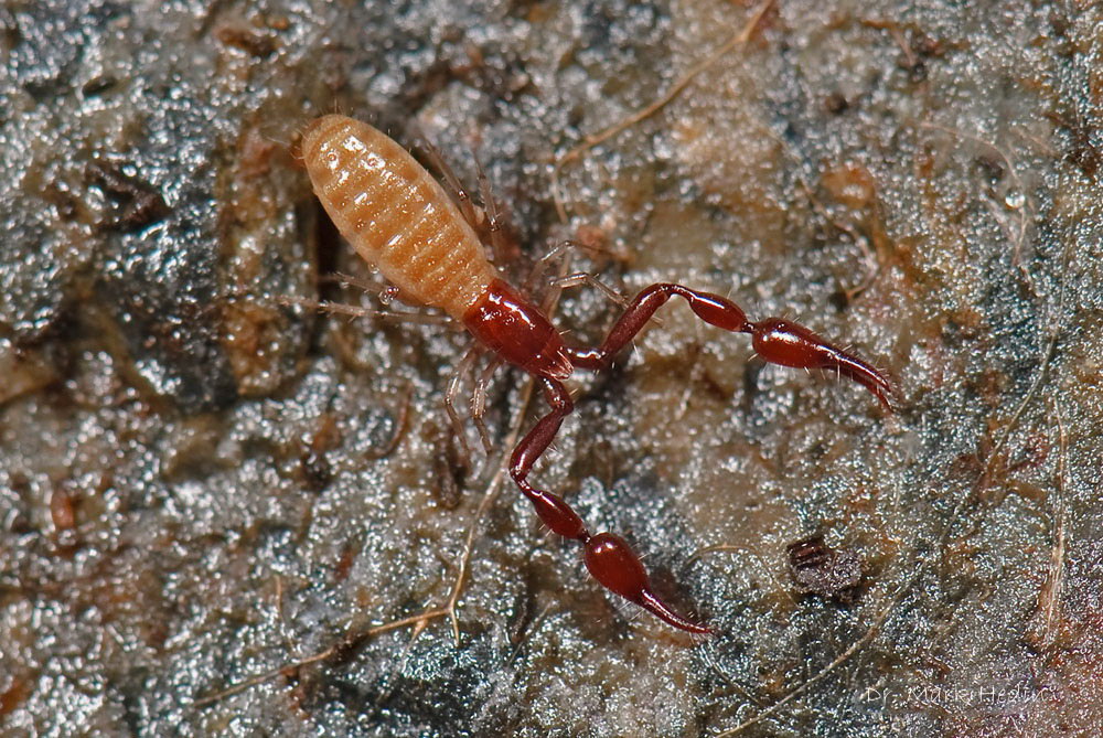
Figure 4: Pseudoscorpion from a conifer wood adjacent to the river Hazel Creek, Great Smokey Mountains, North Carolina. The tergits are not divided in this species. Copyright is held by Dr. Mark Hedin, website: http://marshalhedinlab.com/, photostream: http://www.flickr.com/photos/23660854@N07/.
The openings of the respiratory organs (spiracles) are situated at the 3. and 4. segment of the ventral surface of the opisthosoma and consist of four short tracheal trunks. Pseudoscorpions do not possess book lungs and no respiratory movement could be observed (Weygoldt, 1969, p17).
The number of eyes differs amongst the different groups of pseudoscorpions. Chthonioidae, Neobisioideae and further possess four lateral eyes (except eyeless troglomorphic species) other pseudoscorpion groups possess only two lateral eyes or none though the lack of eyes in these groups corresponds not obligatory to a troglobiontic lifestyle (Weygoldt, 1969, p19).
Pseudoscorpions exhibit two different feeding modes that are reflected in the different cheliceral morphologies (Weygoldt, 1969, p8-12).
Representatives of the Chthoniidae, Neobisiidae and related families masticate their prey by means of their massive chelicerae whereby digestive fluid is spread over the prey and subsequently is ingested with the digested components. Only a small globule of non-digested material is left over. This feeding mode allows only to feed on relatively small prey in comparison to the size of the predator (Weygoldt, 1969, p8-12).
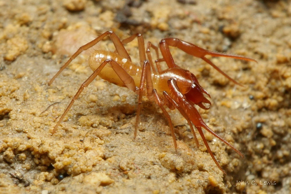
Figure 5: Pseudotyrannochthonius typhlus of the family Chthoniidae. The picture was taken in the caves of Mole Creek, Tasmania. Troglomorphic eyeless pseudoscorpion (6 mm body length) that masticates its prey with its massive chelicerae. Pseudoscorpions of the family Chthoniidae do not possess venom glands. This condition seems to be plesiomorphic and assumingly is not due to a secondary loss (Murienne et al., 2008). Copyright is held by John C. Douglas, Website: http://www.tasmanianspiders.info/.
Pseudoscorpions of the family Olpiidae, Garypidae, Feaellidae and others as well as representatives of the suborder Cheliferoidea exhibit another feeding mode, that allows to extend the prey spectrum to larger animals which could not be crushed with the chelicerae or carried around. Representatives of this group possess small chelicerae and a specialized preoral chamber, that together function as an “injection syringue” (Weygoldt, 1969, p8-12). A small hole is torn in the body wall of the prey whereupon digestive fluid is injected and later reabsorbed together with digested prey compounds. The body wall of the same prey can be pierced several times at different body parts. Usually the complete cuticula of the prey stays nearly intact after the pseudoscorpion has fed (Weygoldt, 1969, p8-12).
The reproduction in pseudoscorpions occurs by way of indirect sperm transfer via a spermatophore (Schaller, 1965; Weygoldt, 1969, p39).
The methods how the females come in contact with the spermatophores differ highly in different representatives of pseudoscorpions (Weygoldt, 1969, p39).
In some species mating does not occur and the male and female act independently in time and space (Weygoldt, 1969, p39). The male deposits spermatophores in the absence of a female and the latter come in contact with the spermatophores more or less by chance. This method leads to “wastage” of sperm cells since many spermatophores are not found or are too old when they are. This mode of sperm transfer furthermore requires a humid habitat, because otherwise the sperm packages would dry out too quickly and a very active lifestyle of the species (Weygoldt, 1969, p40-42).
Other species also do not mate but the male only produces a spermatophore in the presence of a female (Weygoldt, 1969, p39).
In the species Serianus carolinensis (Olpiidae) males possess additional opisthosomal silk glands, with which signal threads can be produced that lead the female to the spermatophore (Weygoldt, 1969, p42-44).
Finally many species exhibit a mating ritual at which both mates actively absolve a “mating dance” whereby this can occur with permanent bodily contact throughout the mating or with bodily contact occurring only at the initiation and the end, where the male assists the female taking up the sperm package from the spermatophore (Weygoldt, 1969, p39, p53-58).
The spermatophores of pseudoscorpions exhibit different grades of complexity where the less complex forms occur in species that do not mate (Weygoldt, 1969, p58).
Pseudoscorpions are oviparous. The females carry the fertilized eggs in a brood sac (Fig. 6) that is attached to the genital area due to a secretion of accessory glands of the genital apparatus. The brood sac is in luminal contact with the genital atrium. The eggs are small and poor in yolk. The embryos are nourished by a nutritive fluid that is secreted from cells in the ovary of the mother (Weygoldt, 1969, p66).
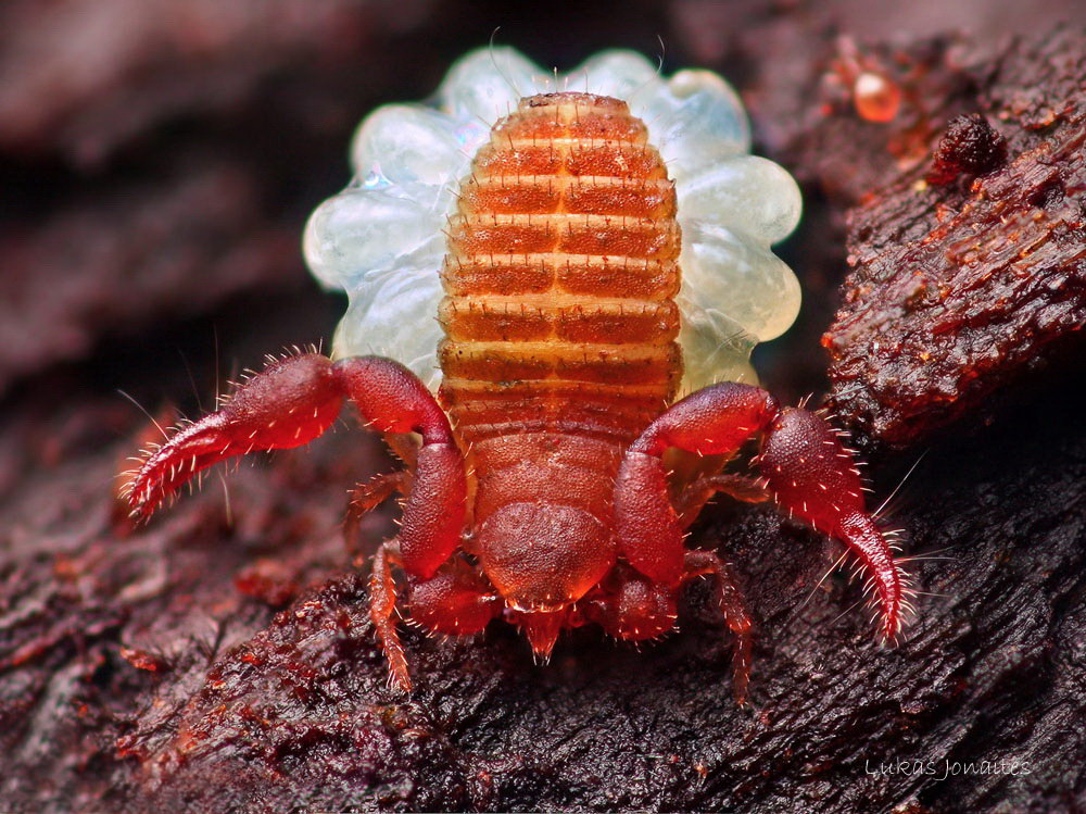
Figure 6: Female pseudoscorpion of the suborder Cheliferoidea with brood sac. This specimen was photographed in Lithuania and has a body size of 2-4 mm. The embryos that are situated in the brood sac, that is attached to the ventral surface of the opisthosma, are inflated by the fast uptake of nutritive fluid with the pumping organ (Weygoldt, 1969, p76-77). The tergits of this species are divided. The chelicerae are small and are utilized as injection syringue (Weygoldt, 1969, p8-12). Copyright is held by Lukas Jonaites, photostream: http://www.flickr.com/photos/38628972@N05/with/5086203158/.
The embryos develop a unique pumping organ that allows them to take up nutritive fluid (Weygoldt, 1969, p91-101).
At the end of the embryonic phase the young molt into the protonymphal state (the first of three juvenile postembryonic states) and leave the mother (Weygoldt, 1969, p68, p102).
Many pseudoscorpions build nests out of threads that are built with the aid of the silk glands, whose efferent ducts open in the chelicerae. Nests are built for molting, hibernation and brooding (Weygoldt, 1969, p24-25).
Many pseudoscorpion species show phoretic behavior (Phoresy: The use of other species for transport) and can be found attached with one chela to the legs of flies (Fig. 7), harvestmen and other animals. As a result of this behavior pseudoscorpions can reach distant areas for what they could not be capable of their own. In Europe the genera Lamprochernes and Pselanochernes are transported by this way.
Phoresy in pseudoscorpions was mainly observed in the families Chernetidae and Cheliferidae but never in the families Chthoniidae und Neobisiidae (Weygoldt, 1969, 116-117).
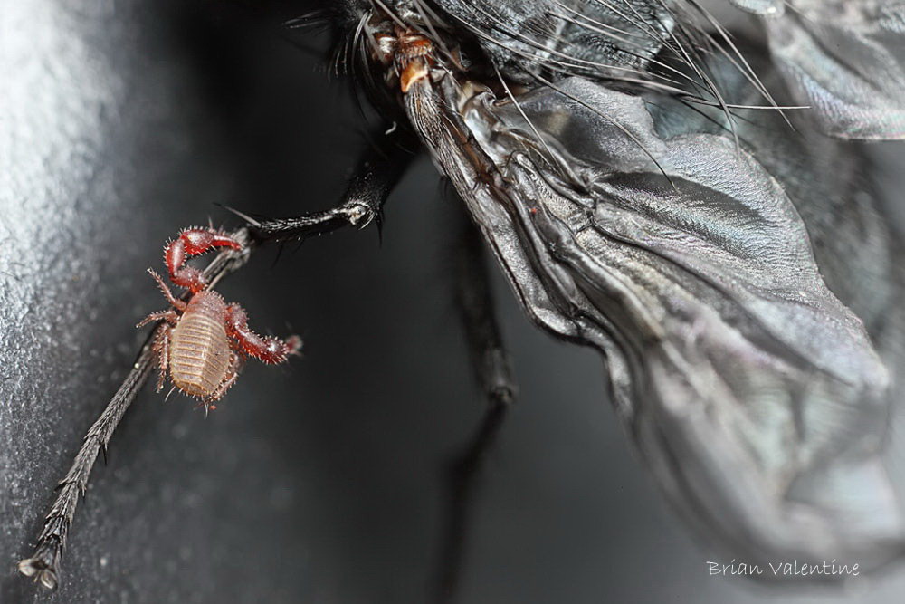
Figure 7: Pseudoscorpion, probably of the suborder Cheliferoidea, attached to the leg of a fly. Some pseudoscorpions “hitch rides“ on other animals on which they hold firmly with aid of their chelae and thereby can surpass great distances. This behavior is termed phoresy (Weygoldt, 1969, 116-117). Copyright is held by Brian Valentine, photostream: http://www.flickr.com/photos/lordv/.
Pseudoscorpions are regarded as being the sister taxon of camel spiders (Solifugae) that together form the clade of the Haplocnemata (Dunlop, 2000; Dunlop, 2010; Shultz, 2007).
Unifying characteristics are the two-segmented chelicerae with a ventral insertion of the movable finger, the occurrence of a “lip” (epistomal-labral plate) situated to the mouth dorsally, elongated patellae of the walking legs, adhesive pulvilli on the tarsi and tracheal openings on the 3. and 4. segment of the opisthosoma. However such a “lip” and this arrangement of the chelicerae can also be found in some mites (Dunlop, 2000; Dunlop, 2010; Shultz, 2007).
After Alberti & Peretti (2002) the morphology of the male genitals and the spermatozoa contradicts the grouping of the Haplocnemata, whereby shared characteristics concerning these findings can be found between the Soifugae and actinotrichid mites.
References
Alberti, G., Peretti, A. V. (2002). Fine structure of male genital system and sperm in Solifugae does not support a sister-group relationship with Pseudoscorpiones (Arachnida). Journal of Arachnology, 30, 268-274.
Dunlop, J. A. (2000). The epistomo-labral plate and lateral lips in solifuges, pseudoscorpiones and mites. Ekológia (Bratislava), 19, 67-78.
Dunlop, J. A. (2010). Geological history and phylogeny of Chelicerata. Arthropod structure & development, 39, 124-142.
Harvey, M., S. (1992). The phylogeny and systematic of the Pseudoscorpionida (Chelicerata: Arachnida). Invertebrate Taxonomy, 6, 1373-1435.
Harvey, M. S. (2002) The neglected cousins, what do we know about the smaller arachnid orders? The Journal of Arachnology, 30, 357-372.
Harvey, M. S. (2007). The smaller arachnid orders: diversity, descriptions and distributions from Linnaeus to the present (1758-2007). Zootaxa, 1668, 363-380.
Harvey, M. S. (2009). Pseudoscorpions of the World, version 1.2. Western Australian Museum, Perth. Available from: http://wamuseum.com.au/arachnids /pseudoscorpions/.
Murienne, J., Harvey, M. S., & Giribet, G. (2008). First molecular phylogeny of the major clades of Pseudoscorpiones (Arthropoda: Chelicerata). Molecular Phylogenetics and Evolution, 49, 170-184.
Schaller, F. (1965). “Mating behavior of lower terrestrial arthropods from the phylogenetic point of view,”. Proc. 12th Intern. Congr. Entomol. Lodon 1964, 297-298.
Schawaller, W., Shear, W., & Bonamo, P. M. (1991). The first Palaeozoic Pseudoscorpions (Arachnida, Pseudscorpionida). American Museum novitiates, 3009.
Shultz, J. W. (2007). A phylogenetic analysis of the arachnid orders based on morphological characters. Zoological Journal of the Linnean Society, 150, 221-265.
Weygoldt, P. (1969). The Biology of Pseudoscorpions. Harvard University Press, Cambridge, Massachusetts.
F. Schramm, authored 2011